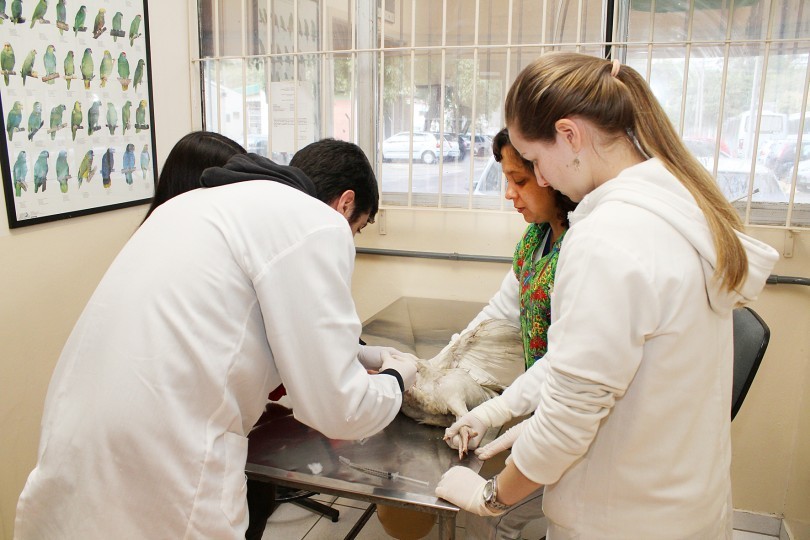
Small animal imaging
That radiation can be harmful if your dog is repeatedly and extensively exposed. That stated, pet insurance would most likely not help cowl the costs of your dog’s x-rays whether it is associated to a pre-existing situation, or in case your plan’s related waiting durations weren’t up but. Positron emission tomography (PET) is a specialised radiology procedure that uses molecular imaging to trace and hint each normal and irregular situations. PET is probably the most advanced nuclear imaging technique, allowing for high-resolution imaging and absolute quantification of biologic mechanisms. PET imaging can be used to comply with the progress of sufferers present process treatment. While PET is mostly used in the fields of neurology, oncology and cardiology, applications in different fields are at present being studied. Compared with an X-ray or magnetic resonance imaging (MRI) scan, a PET/CT enables doctors to examine medical situations and abnormalities at a mobile level.
Y comparta sus puntos de vista y vivencias con otros pacientes, familias y cuidadores. El trámite es totalmente indoloro e inofensivo y tarda aproximadamente entre 10 y 15 minutos. Se anestesia la parte de atrás de la garganta y se introduce a través de esta un catéter largo, maleable pero estable (llamado "sonda") el cual tiene en su radical final un transductor de ultrasonido. El Departamento de Cardiología de la Clínica colabora con los Departamentos de Radiología y de Cirugía Cardiaca para lograr un diagnóstico veloz y preciso del paciente. El médico que solicita la prueba le indicará si debe tomar la medicación frecuente o ésta debe ser suspendida previamente a la realización del test.
Lo que se siente durante el examen
La finalidad de este blog es proporcionar información de salud que, en ningún caso reemplaza la solicitud con su médico. El paciente se desviste de cintura para arriba y se tumba en una camilla, de lado izquierdo con el brazo izquierdo bajo la cabeza. Con el transductor en distintas ubicaciones, adquirimos las diferentes imágenes del corazón. En líneas generales, con el electrocardiograma valoramos la actividad eléctrica, y el ecocardiograma nos deja valorar la estructura y función cardiaca. Los exámenes de detección temprana de enfermedades cardiacas nos dejan detectar inconvenientes en el corazón y su desempeño antes de que aparezcan los síntomas. El propósito de la detección temprana es detectar una enfermedad en su etapa más temprana y mucho más tratable.
 The integration of expertise in echocardiogram interpretation holds super potential for improving diagnostic accuracy, effectivity, and patient outcomes. As these applied sciences continue to evolve, clinicians should embrace their advantages and adapt their practice to leverage the advantages provided by these innovative options. Telemedicine platforms enable remote interpretation of echocardiogram studies, allowing clinicians to entry and interpret research from completely different places. This technology facilitates collaboration between consultants, allows well timed interpretations, and improves entry to specialised care.
Risks
Your health care provider can use the images from the test to find heart illness and other coronary heart conditions. An echocardiogram—also often identified as an echo, cardiac echo, or cardiac ultrasound—allows healthcare providers to see the guts's structure and blood circulate. Providers can observe the rhythm of a coronary heart and measure the size and performance of the heart’s chambers and valves. The echocardiogram is an ultrasound scan of the heart that reveals shifting footage that present the structure and performance of the heart. It reveals correct information on the guts pumping operate and heart chamber sizes.
Heart conditions diagnosed with echocardiograms
Moreover, it might possibly additionally pick up on irritation, heart murmurs, congenital disease, and the like. Although the concept of an echo exam is frightening, in actuality, it is nothing to be afraid of. If anything, echocardiograms are identical to an ultrasound, but on your heart. Ultimately, an echocardiogram converts sound waves into images. It is then these pictures that capture your coronary heart and Https://Staal-Carson.Mdwrite.Net/Raio-X-24-Horas-A-Importancia-Do-Diagnostico-Rapido-Para-A-Saude-Do-Seu-Cao its neighboring blood vessels. An echocardiogram is usually step one when trying to identify heart illness.
If Your Echo Result Is Normal, Does That Mean Your Heart Is OK?
The hairs shall be separated with a small amount of alcohol, and ultrasound gel might be utilized to the area to assist improve the contact between the probe and your canine's body. Most dogs do not require shaving, but in some cases, shaving a small patch on either side of the chest is necessary to assist optimize the standard of the photographs. If your canine does not require any sedation before their echocardiogram, they'll eat and drink normally. Knowing the standing of your dog's coronary heart can even help your veterinarian determine if it is potential to deal with other sicknesses extra aggressively (for example, kidney disease or cancer). MyHeart is a group of physicians devoted to empowering sufferers to take management of their well being. Read by over 1,000,000 folks yearly, MyHeart is shortly becoming a "go to" useful resource for sufferers across the world.
Different types of echocardiograms
A well being specialist will follow tips for amassing several varieties of pictures and data. An aneurysm is a widening and weakening of a part of the heart muscle or the aorta (the massive artery that carries oxygenated blood out of the center to the remainder of the body). Shunts could be seen in atrial and ventricular septal defects but additionally when irregular blood move is pushed by way of the circulation from the lungs and liver. When your coronary heart is beneath stress, your sonographer can see particulars they received't be able to see should you were lying on the exam table. These embody problems along with your coronary arteries or the lining of your heart. This test exhibits how nicely your heart can face up to activity.
STEMI Management Guide: From Timely Diagnosis to Effective Intervention
Taking an active position in your prognosis and care might help you feel comfortable with every step of the method. Ask your provider when and tips on how to take your usual medications. You may have to avoid taking sure coronary heart medications on the day of your take a look at. You may also need to alter your dose of diabetes medication. An echocardiogram and an electrocardiogram (called an EKG or ECG) both verify your coronary heart.
What Does An Echocardiogram Show – Patterns Of Blood Flow
In such cases, extra imaging modalities (e.g., transesophageal echocardiography) or alternative diagnostic checks could additionally be necessary to obtain a clearer assessment. The echocardiogram report evaluates the size and function of the cardiac chambers, including the left ventricle, proper ventricle, left atrium, and proper atrium. Volume measurements, similar to left ventricular end-diastolic quantity (LVEDV) and left ventricular end-systolic volume (LVESV), provide a more accurate assessment of chamber size. An echocardiogram, also known as an echo, is a non-invasive ultrasound requiring specialised tools, coaching, and knowledge. It is used by veterinary cardiologists as a diagnostic tool to judge the center. It allows them to discover out the heart's dimension, shape, and performance, and discover its chambers, valves, and different surrounding structures. It also evaluates the most important blood vessels that go away the center.


