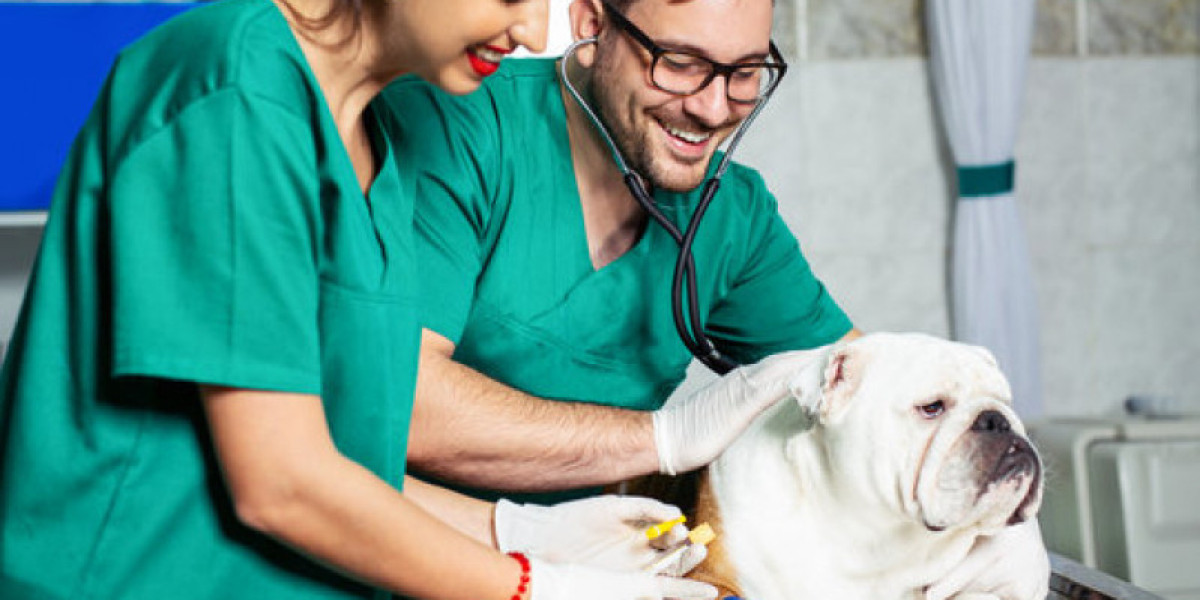
Why Would My Pet Need an Echocardiogram?
As talked about above, your native vet will only be in a position to perform this check a number of occasions a month if they don’t have the suitable tools. There are a variety of opinions on how often echocardiograms must be ordered. In general, physicians are probably to order more echocardiograms than are necessary. Recently, Dr. Catherine Otto[1] supplied what seem like acceptable guidelines for Veuarc.Com the usage of echocardiography in sufferers with mitral regurgitation.
Are you concerned about your pet?
Ultrasound examinations can be used to examine the heart, stomach organs, eyes and reproductive organs in canine. Other organs, similar to those in the stomach, are rarely examined during an echocardiogram. Since the test is painless, non-invasive, and generally takes now not than fifteen minutes, your canine will not require any sedation or anesthesia. However, delicate sedation may be needed for canines who are very fearful or anxious as a result of they want to stay utterly still through the testing to get clear pictures and essentially the most accurate analysis and diagnosis.
La conclusión de este estudio es que múltiples cambiantes mensurables mediante ecocardiografía Doppler pueden ser usadas para la predicción de la insuficiencia cardiaca en perros con valvulopatía mitral crónica o cardiomiopatía dilatada.
Es fundamentalmente imposible valorar las radiografías sin un corte preexistente a resultas del conocimiento del historial, los descubrimientos de la exploración física y los desenlaces de laboratorio de exames veterinarios realizados previamente.
If you live in an area with the next common price of residing, you'll most likely additionally pay extra for x-rays (and different veterinary medicine services) than you'll pay in an space with a decrease price of living.
 "The most typical coronary heart disease in canines, representing seventy five percent of all coronary heart illness, is characterised by degeneration of the mitral valve (the valve between the left atrium and left ventricle)," Gordon says. According to Gordon, this degeneration results in a leaky valve, which causes the canine's heart chambers to enlarge. "It eventually can result in coronary heart failure in some canines. It additionally causes a coronary heart murmur that can be detected by your vet," Gordon says. Although there is no treatment for congestive coronary heart failure in canines, many can stay fortunately for months and even years with the proper treatment.
 They involve the injection of gallium citrate, a radioactive tracer. Gallium scans are a multiday course of and are sometimes carried out 1 to three days after the tracer is administered. This is because healthy heart tissue tends to take in more of the tracer than unhealthy tissue or tissue that has decreased blood move. However, these scans should be read rigorously and defined by a doctor, as it’s potential for noncancerous conditions to look like cancer on a scan.
PET plus CT
While attenuation-corrected pictures are typically more devoted representations, the correction course of is itself susceptible to significant artifacts. As a end result, each corrected and uncorrected photographs are at all times reconstructed and skim collectively. FDG is probably the most generally used tracer for imaging muscles, and NaF-F18 is probably the most extensively used tracer for imaging bones. If you are afraid of enclosed areas, you might really feel some nervousness whereas in the scanner. Be certain to tell the nurse or technologist about any anxiousness inflicting you discomfort. The PET-CT or PET-MRI scanner is a big machine that appears slightly like a large doughnut standing upright, much like CT or MRI scanners.
En el presente informe se optó por una sonda lineal, puesto que se prefirió una frecuencia mucho más alta para la caracterización de las consolidaciones identificadas. Con base en las recomendaciones de la medicina humana, se puede elegir la sonda lineal para la evaluación de la consolidación pleural y subpleural (25, 40). Sin embargo, debe tenerse presente que la sonda microconvexa de forma frecuente se recomienda para el análisis vertical de artefactos de la área pulmonar. Un macho Maltipoo de 1,5 años fue remitido al servicio de emergencias por disnea, debilidad, tos y anorexia de 3 días de evolución. A la exploración física, el perro presentaba taquipneico (40 respiraciones/min) con marcado esfuerzo respiratorio, tenía restallantes cráneo-dorsales locales auscultados bilateralmente y temperatura corporal rectal de 36,6 °C. En la auscultación cardíaca se observó un soplo cardiaco sistólico apical derecho (grado 1/6). La ecografía torácica en el punto de atención efectuada por el médico de emergencias que lo atendió identificó caudodorsalmente las vías B difusas.


