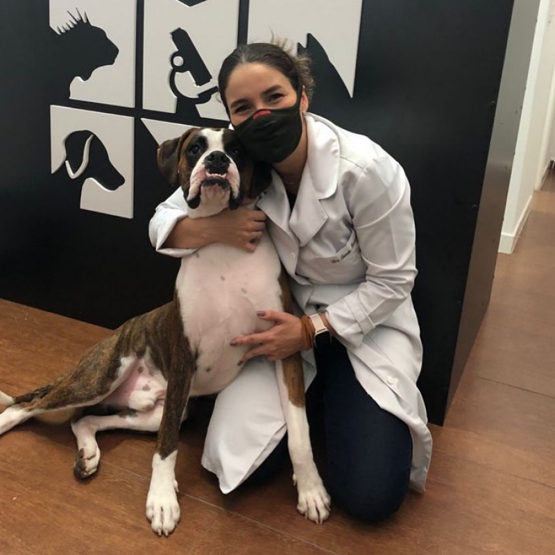
Hospital Veterinario Vetpro Provença
"Tanto el dueño como el clínico reconocen que estos dos causantes son los que llevan a decidir a qué centro laboratório veterinario são paulo acudir", afirma Llamas. En relación a la composición del ámbito, "nos encontramos en un instante de cambio en el campo de las clínicas veterinarias de pequeños animales", afirman desde AMVAC. Nuestras instalaciones y equipamiento de vanguardia nos dejan ofrecer a tus mascotas el precaución que se merecen. Contamos con un equipo de expertos cualificados y expertos, instalaciones modernas y pertrechadas, y una amplia gama de servicios para agradar todas y cada una de las pretensiones de tu mascota. Los guantes plomados permiten inmovilizar de forma manual al tolerante mientras que se realiza el estudio radiológico.
 Summer is a season of enjoyable and adventure, but it additionally brings along scorching temperatures that may pose severe risks to our pets. By being vigilant and knowledgeable, you can make a significant distinction in your pet's health and potentially save their life. A detailed description of arrhythmia therapy is beyond the scope of this article; nonetheless, some basic information is warranted. The choice of whether or not an arrythmia warrants therapy is dependent upon the hemodynamic consequences, the probability of an increase in severity, and the dangers posed by medicine.
Why Trust CVCA for Veterinary Cardiology?
AF is often current with heart failure; subsequently, sufferers typically show indicators of left-sided congestive coronary heart failure and are normally train intolerant. The complexes look irregular as a outcome of the ventricles prematurely fired before the sinus advanced started, meaning the impulse needed to depolarise the myocardium cell by cell. This takes longer, and due to this fact the complicated seems broad and bizarre. It could have all of the doctor’s findings and fala sobre isso recommendations, intimately. A full report shall be faxed over the same day to your major care veterinarian, along with some other veterinarians who are concerned in your pet’s care.
Some canine breeds are at particular threat for coronary heart illness, so your veterinarian will always wish to check these canines and can doubtless additionally recommend a examine if you’re considering breeding. This info may help your pet’s veterinary cardiologist ensure that your pet’s heart is working properly. Symptoms of heart illness often take time to be noticeable to pet homeowners. An echocardiogram will provide a piece of thoughts that you simply and your veterinary heart specialist can provide your pet the support and care they want. The P wave indicates atrial depolarization, the QRS complicated indicates ventricular depolarization, and the T wave indicates ventricular repolarization.
What happens during your pet’s echocardiogram procedure?
This certification is awarded by Sonopath to sonographers demonstrating superior level sonographic image capturing and efficiency. When deciding whether or not to buy any sort of insurance coverage or low cost plan, it's critical to make an knowledgeable determination about the sort of protection you want. Some of those situations might embrace elevated travel and day with out work from work, which might increase your fuel price range and affect your bills. The information is current and up-to-date in accordance with the latest veterinarian research. Advanced Imaging consists of MRI – Magnetic Resonance Imaging and CT – Computed Tomography – are each non-invasive, extremely correct diagnositc instruments for unwell or injured pets. 5,534 pet homeowners requested and acquired a free no-obligation quote from one of the above corporations within the final 30 days. All price ranges are calculated as averages from various 2023 stories, together with CareCredit Canine Journal, PetKeen and Pawlicy Advisor.
 Depending on the symptoms, your vet might suggest magnetic resonance imaging (an MRI) or a CT scan. These are high-definition imaging methods that can pinpoint small issues within your pet that could be tougher to find or diagnose with an x-ray. However, X-rays will not be efficient in identifying extra delicate or non-specific stomach issues, such as inflammation, infections, or sure forms of cancer. If the dog needs to be sedated or anesthetized for the X-ray, it may possibly take longer as there shall be further time wanted for the sedation to take effect and for the dog to get well afterward.
Detailed Imaging Results
There is a "blind spot" ventrally and centrally along the cardiac silhouette on the VD/DV images the place pulmonary lesions will not be seen. Recumbent lesions (lesions in the down lung when the patient is in lateral recumbency) will border efface with the encircling pulmonary parenchyma from atelectasis. For emergency circumstances, a dorsoventral thoracic radiograph can be carried out initially but ought to be followed up with a whole set of thoracic radiographs (right lateral, left lateral, and VD/DV) after the affected person has been stabilized. The pleural space exists between every lung lobe at the interlobar fissure as properly as across the lung lobes themselves. The pleural space reflects again on itself on the mediastinum.1,2 These fissures are not seen on regular thoracic radiographs except instantly tangential to the x-ray beam. The commonest pleural fissure noted is the one between the right center and right caudal lung lobes on a left lateral radiograph (FIGURE 2). During your pet’s Sick Visit, your veterinarian may suggest radiology services to get a better have a look at your pet’s bones, joints, or different inside constructions.


